Retinal Disease
Retinal Disease – AMD
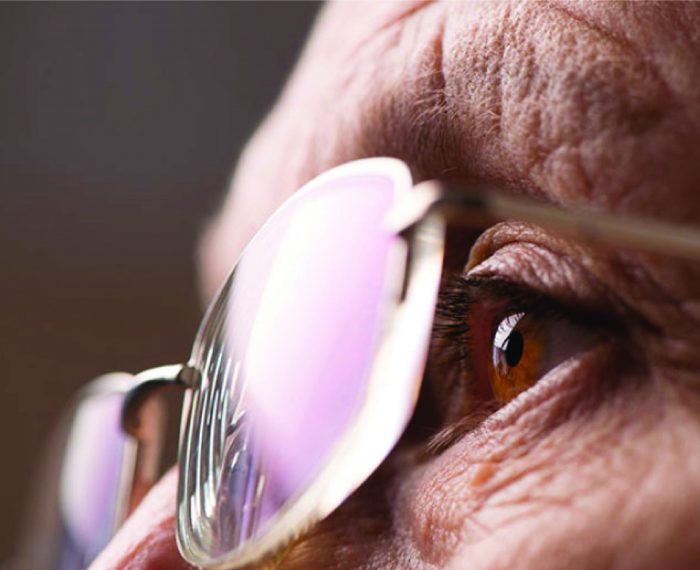
The retina is the inner most layer of the eye and is the imaging centre.
Located at the back where the optic nerve is, the retina is responsible for turning light into nerve signals from the back wall of the eye to the area of the brain responsible for sight.
Conditions that can affect your retina (e.g. ageing, chronic diseases like diabetes and trauma to the eye) may lead to vision loss.
THONEH’s Consultant Ophthalmologists are well experienced in treating various retinal conditions to prevent vision loss. They are experts in their field using the latest technologies and best practices for diagnosis and management of these conditions.
AGE-RELATED MACULAR DEGENERATION ( AMD )
Introduction

Age-related Macular Degeneration (AMD) is a chronic eye disease and one of the leading causes of severe vision loss in people 60 years of age and above. It is also one of the leading causes of adult blindness in the world.
Contrary to what many believe, vision loss – and specifically AMD – does not have to be an inevitable consequence of ageing. Protect your vision by being active in your healthcare. Visit your eye doctor regularly.
What is AMD?
AMD is a disease that can affect the sharp central vision needed for “straight-ahead” activities like reading, driving, telling time and recognize faces.
Sometimes AMD advances so slowly that you notice little change in vision. Sometimes AMD progresses quickly, resulting in rapid vision loss. AMD causes no pain, but can rob you of you ability to see what is on front of you. There are two types of AMD: Dry & Wet.
Dry AMD
In dry AMD, which makes up to about 85% of all AMD cases, the light-sensitive cells in the retina die, affecting “straight-ahead” vision. The most common symptom of dry AMD is blurred vision. Dry AMD tend to develop slowly, but may develop into a more severe form of AMD called wet AMD.
Wet AMD
Wet AMD, accounts for about 15% of all AMD cases, has abnormal blood vessels growing under the macula. This may lead to bleeding, scar formation and permanent damage. Damage occurs more rapidly than in the dry form and tends to lead to more severe loss of central or “straight-ahead” vision. If detected in time, relevant treatments may stop the progression of vision loss.
Am I at Risk?
The two greatest risk factors for developing AMD are:
- Increasing age – about 25% of people over 65 years of age have AMD.
- Having AMD in one eye – of those with AMD in one eye, about 40% wil develop AMD in the other eye within five years.
Other risk factors for developing AMD include:
- Smoking
- Race – Caucasians appears to have higher risk
- A family history of the disease
- Low dietary intake of certain vitamins and minerals
- Gender – women appear to be at greater risk than man
Am I Seeing These Symptoms?
Symptoms of AMD include:
- Decreased Visual Acuity
- Dim, or less sharp central or “straight-ahead” vision
- Loss Of Contrast Sensitivity
- Colors that seem washed out or dull
- Central Scotoma
- A blank or blind spot in your central vision
- Metamorphopsia
- Seeing objects as wavy or curved.
How is WET AMD Diagnosed?
- Fundus Photography
- Fluorescein Angiography. This is a test which provides information about the blood circulation in the retina that cannot be seen by routine eye examinations. The Fluorescein dye is injected into the blood via a vein in the arm and rapidly reaches the eye. The dye circulates through the retina and highlights any abnormalities or damage. The abnormal new blood vessels seen in Wet AMD have weak, fragile walls and the dye leaks through them, outlining them clearly.
- Indocyanine Green Angiography. Indocyanine Green (ICG) Angiography has different properties to Fluorescein and highlights the deeper layer of the retina and the choroidal circulation (the source of the abnormal blood vessels), which is normally hidden from view. It enables different types of new vessels to be identified.
- Optical Coherence Tomography. Optical Coherence Tomography (OCT) is a non-invasive diagnostic imaging technique that uses light to produce very high-resolution cross-sectional images of the tissue layers within the retina. These layers at the macula can then be studied and measured in microscopic detail. By comparing the structure and the thickness of the layers measured by the OCT against a normal healthy retina, eye specialists can detect any Wet AMD even at a very early stage.
- Optical Coherence Tomography Angiography. Optical Coherence Tomography Angiography (OCTA) is a fast, new imaging modality, which uses the principles of OCT to define the retinal vascular structure, using sequential B-scan to detect blood flow. En face images can then be generated showing the superficial and deep retinal capillary plexus and the choroid. Cross-sectional OCT combined with depiction of flow can help demonstrate the location of the vascular abnormality. Although OCTA detects neovascular AMD by detecting flow in a vascular complex, it does not show leakage.
How can AMD be Treated?
AMD causes permanent vision loss. Successful treatment can stabilize and /or slow vision loss or in some cases, restore vision. Although new treatments are always being researched, treatment options today are:
- Anti-VEGF Injection. The new blood vessels are prompted to grow by a protein called Vascular Endothelial Growth Factor (VEGF). Anti-VEGF drug is injected into the eye to block the protein responsible for the growth of new blood vessels. Anti-VEGF drug is injected into the cavity where it can spread to the retina. The injections are generally administered at four to six week intervals.
- Photodynamic therapy, used to help treat wet AMD. In this procedure, a light-activated drug is injected into the blood stream and travels to the abnormal blood vessels in the eyes. It is then activated in the eye by a non-heat laser to help reduce the risk of further vision loss. PDT is no longer used as a monotherapy as patients continue to lose vision in the first six months. If used in conjunction with Anti-VEGF this vision loss can be prevented. There is one type of new vessels called polypoidal vascular choroidapathy for which this combination may be preferable to giving Anti-VEGF by itself.
- Laser photocoagulation. In this procedure, a high-energy beam of light is used to destroy leaky blood vessels, preventing further loss of vision. Through heat, the treatment damages the retina at the treatment spot, producing scars. Therefore it should only be used for treating new vessels that are not under the central macula. This treatment is only for a small percentage of patients with a particular type of AMD.
- Optical Aids and Lights. Low vision optical aids often improve vision for people with macular degeneration. Many different types of magnifying devices are available. Spectacles, hand or stand magnifiers, telescope, and closed circuit television for viewing objects are some of the available resources. Aids are prescribed by your ophthalmologist or by referral to a low vision center. Bright illumination properly directed for reading and close work are often beneficial. Special lamps can also be helpful. Books, newspapers, and other items available in large print offer further help. A patient with macular degeneration can be helped. Fortunately, visual aids are available to assist many patients in leading a comfortable and relatively normal life. With these devices and proper motivation, people with visual loss can often read, do modified close-up work, and continue to take care of themselves.
- If you are over the age of 50, or if your family has a history of retinal problems, you should have your eyes checked periodically for signs of eye problems like macular degeneration. Early detection and subsequent treatment if indicated, may help prevent additional visual loss. If you have additional questions or would like any further informations, contact your ophthalmologist.